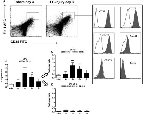
Figure 3.
EPCs detected in the kidney mainly express surface markers of ECFC. CD34+/Flk-1 positive cells (measured using flow cytometry) were increased 3 days following endothelial selective injury (A, B). The major part of CD34+/Flk1+ cells express endothelial and lack hematopoietic surface markers (A). ECFCs (C) represented the major and EC-CFUs (D) the minor type of EPC in the murine kidney following selective EC injury.

