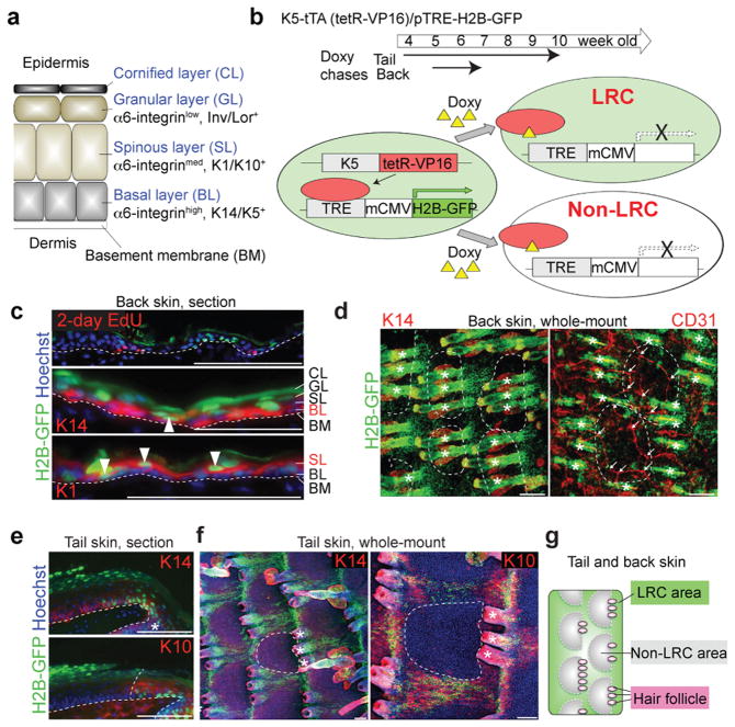
Figure 1. H2B-GFP LRCs and non-LRCs reside in distinct territories of the epidermis.
a, Structure of mouse inter-follicular epidermis and associated markers. b, Scheme of the strategy to detect infrequently dividing cells as H2B-GFP LRCs in adult skin of transgenic mice. c-i, Immunostaining of back and tail skin after H2B-GFP pulse-chase is shown in 10 μm tissue sections or whole mount using antibodies to differentiated markers, as indicated. CD31 is a vascular marker. Hoechst is a DNA-specific stain. The dashed line surrounds non-LRC areas (d,e,h,i) or represents epidermal-dermal junctions (c,f,g). Arrowheads indicate LRCs in the K14+ BL (middle) or K1+ SL (bottom) (c). (e) Arrows indicate branch points of blood vessels. Asterisks indicate HFs. Scale bars, 100 μm (c-i). j, Schematic view of the tail and back epidermis indicate LRC and non-LRC areas (territories) organization relative to hair follicles. Experiments are repeated twice with 2 mice for all representative images (b-i).

