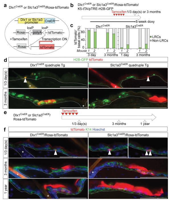
Figure 4. Slc1a3CreER and Dlx1CreER mark long-lived stem cells in the back epidermis.
a, Schematic representation of the tamoxifen (TM)-inducible CreER system. b, Tamoxifen and doxycycline pulse-chase scheme to examine the relationship of Dlx1CreER- and Slc1a3CreER-marked cells with H2B-GFP LRCs in quadruple transgenic mice. c, Fraction of LRCs and non-LRCs in tdTomato+ basal cells in back skin from Dlx1CreER or Slc1a3CreER at 1/3 days or 3 months post-TM. Data have been obtained from 2 mice per condition; data from each mouse are shown as an individual bar. d, Fluorescence images of back skin sections of quadruple-transgenic mice show co-localization of Dlx1CreER, but not Slc1a3CreER, marked cells and generated clones with LRCs. e, Scheme for long-term lineage tracing. f, Lineage tracing in back skin. Arrowheads indicate tdTomato+ basal cells. The dashed line represents epidermal-dermal junctions. The tdTomato signal is extremely intense in the cornified layer (CL), the outermost skin layer, when compared with the basal and suprabasal layers. This is likely because CL contains multiple layers of stacked cells or because the tdTomato reporter is more active there. Interestingly, Slc1a3CreER clones spread away in CL from their BL-marked areas, suggesting that Slc1a3CreER BL cells contribute broadly to the outer skin layer. Asterisks indicate HFs. Scale bars, 100 μm (d, f). Experiments are repeated twice with 2 mice for representative images in (d); and twice with 4 mice (Slc1a3 1 day), 3 mice (Dlx1 3 days and Dlx1 3 months) and 2 mice (Dlx1 1 year, Slc1a3 3 months and Slc1a3 1 year) for images in (f).

