Fig. 4.
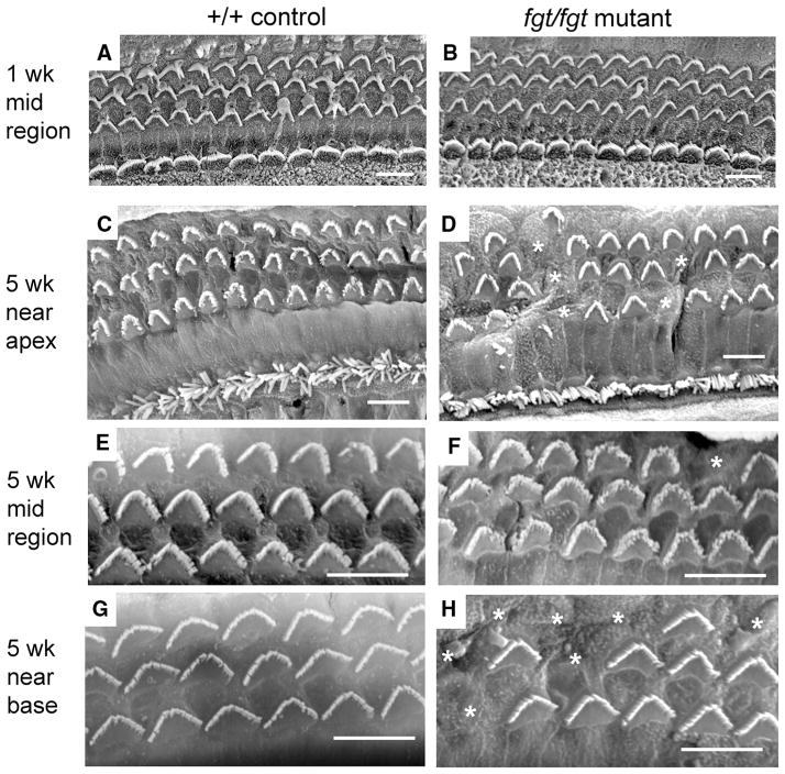
Progressive hair cell loss in Ap1g1fgt mutant mice. SEM was used to examine surface preparations of the organ of Corti in cochleae from +/+ control (a, c, e, g) and fgt/fgt mutant (b, d, f, h) mice at 1 week (a, b) and 5 weeks (c–h) of age. In panels A–D outer hair cells constitute the top three rows and inner hair cells the bottom row, only outer hair cells are shown in panels E–H. At 1 week of age, no differences in outer or inner hair cell bundle morphology or survival were observed between control (a) and mutant (b) cochleae. In mutant cochleae at 5 weeks of age (d, f, h) stereociliary bundles (marked with asterisks) were missing in outer hair cells but not inner hair cells. Loss of outer hair cell bundles was more frequent near the apex (d) and near the base (h) than in the mid region of the cochlea (f). Sporadic regions near the apex also exhibited a disrupted patterning of outer hair cell bundles (left-most part of d). Black scale bars at lower right of each panel represent 10 micrometers

