Figure 1.

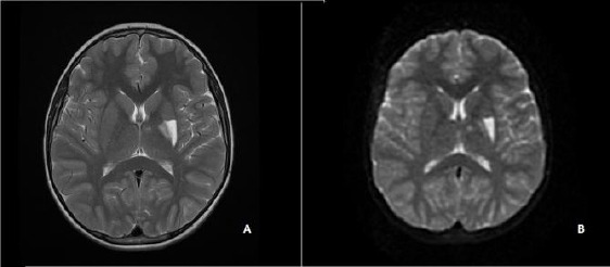
The cerebral MRI showing a hyperintense lesion at the left putamen and globus pallidus interna and externa on T2 (A) and diffusion weighted images (B).

The cerebral MRI showing a hyperintense lesion at the left putamen and globus pallidus interna and externa on T2 (A) and diffusion weighted images (B).