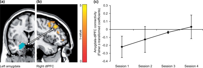
Fig. 4.
Resting-state functional connectivity from left amygdala to right dlPFC was altered in BPD patients over sessions. (A) For illustration, the amygdala mask (blue) is displayed on an anatomical template in axial view (left is left). (B) DlPFC cluster showing a linear functional connectivity increase with left amygdala. Voxels are displayed on a saggital slice (x = 45) of an anatomical template with an uncorrected voxel threshold of P < 0.05 and k > 10 for visualization purposes. Right is anterior. (C) Beta estimates from amygdala-dlPFC connectivity, extracted from the peak voxel (45,23,40). Error bars = SEM.

