
Fig. 5.
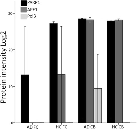
PARP1, APE1 and Polβ protein levels are modified in AD. Quantitative protein detection was performed by mass spectrometry (nLC and Thermo Q Exactive). Levels of base excision repair proteins PARP1, APE1 and Polβ were compared in Alzheimer’s patients (AD) and healthy controls (HC). Reference to house-keeping proteins is shown in Additional file 1: Figure S1. Black bars: PARP1, grey bars: APE1, light grey bars: Polβ. DNA glycosylase OGG1 was below the detection limit. Abbreviations: FC = Frontal cortex, CB = Cerebellum