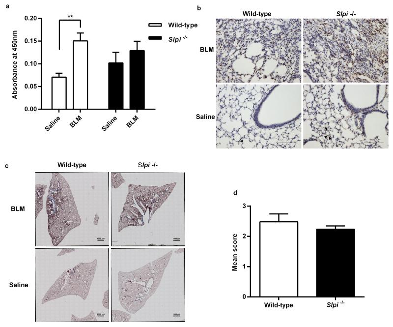
Figure 2. The effect of Slpi deletion on alveolar macrophage TGF-β activation and pulmonary fibrosis.
(a) pSmad2 levels in nuclear extracts from BAL cells from Slpi−/− and wild-type mice 28 days post-bleomycin (BLM). Data expressed as mean absorbance at 450 nm ± SEM; n ≥ 5; **P < 0.005 BLM vs. saline. (b) pSmad2 immunohistochemistry. Representative images captured using original magnification x20, scale bars 100 μm. Arrowheads identify positively stained cells within a single, representative, alveolus in saline treated mice only; there is just one arrowhead in the wild-type mouse and three in the Slpi−/− mouse. (c) Masson’s trichrome staining 28 days post-BLM treatment. Representative, whole lung lobe, cross sections. (d) Quantitative assessment of trichrome staining. Data expressed as mean Ashcroft Score ± SEM; n ≥ 3.

