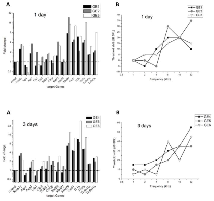
Figure 3.
Changes in target gene expression and ABR threshold shifts in the implanted ear following cochlear implantation for each animal from the 1-day and 3-day groups. The gene expression changes are again grouped in each subplot from left to right as follows: housekeeping gene (column 1), ion homeostasis genes (columns 2–6), tissue remodeling genes (columns 7–11), and inflammation-related genes (columns 12–16).

