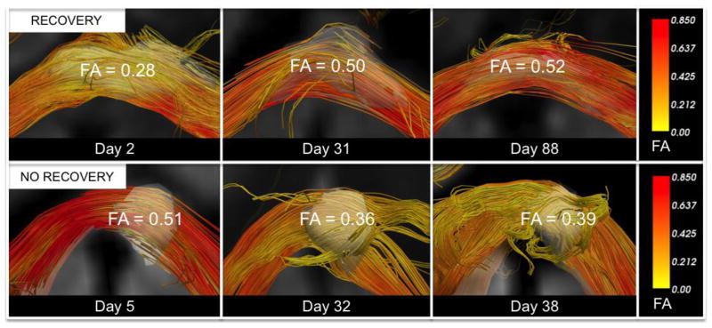
Figure 2. Longitudinal Fractional Anisotropy Changes within White Matter Tracts Affected by Traumatic Axonal Injury.
A traumatic axonal injury (TAI) lesion in the splenium of the corpus callosum of patient 6 (top row) demonstrates FA recovery, as defined by a longitudinal increase in FA that exceeds the coefficient of variation for FA within the splenium of the corpus callosum in the two control datasets. A splenium TAI lesion for patient 7 undergoes a longitudinal decline in FA (bottom row). Each TAI lesion is rendered in 3-dimensions as a semi-transparent white region of interest so that tracts can be visualized passing through the lesions. Fiber tracts were generated using the lesions as seeds. Tracts are color-coded using mean tract FA as a scalar (right panels). All tracts were reconstructed using Diffusion Toolkit version 0.6.2 and TrackVis version 5.2 (Wang & Wedeen, Athinoula A. Martinos Center for Biomedical Imaging, www.trackvis.org).

