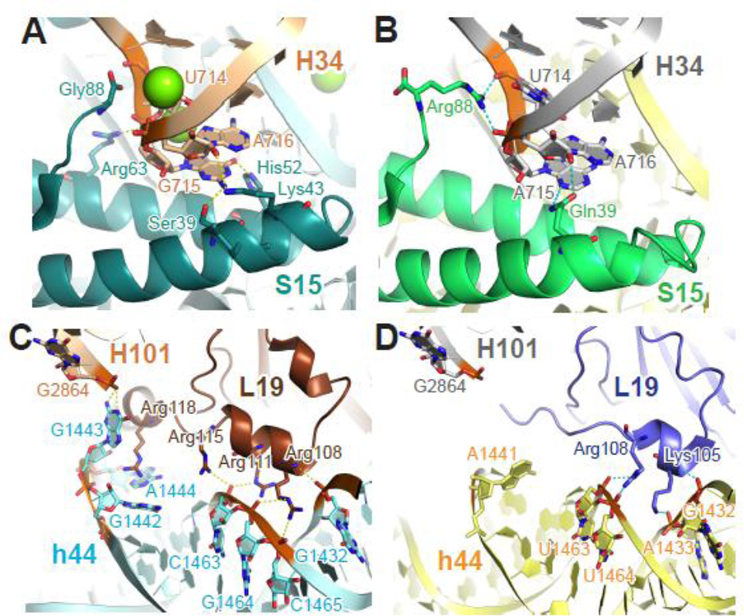
Figure 5. Phylogenetic differences at bridges B4 and B6.
Bridge B4 (A–B) and B6 (C–D) from T. thermophilus (A, C) and E. coli (B, D) ribosomes share the same overall structure despite the phylogenetic differences in protein S15 and L19. In the T. thermophilus B6 (C), additional interactions form due to the unique C-terminal extension of L19 and a three-nucleotide bulge (nt 1442–1444) of h44. Teal (T. thermophilus), green (E. coli), small subunit proteins; brown (T. thermophilus), slate (E. coli), large subunit proteins; otherwise color scheme is the same as Figure 4. Panel A and C were generated using PDB entries 2WDK and 2WDL 39, and panels B and D were generated using PDB entries 3R8O and 3R8T 24.

