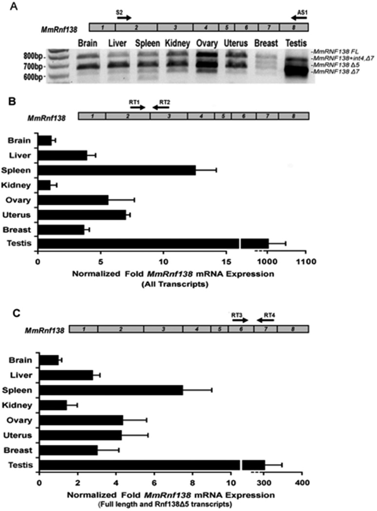
Fig. 3.
RT-PCR analysis of Mus musculus Rnf138 alternative transcripts. (A) RT-PCR for Rnf138 was performed from eight tissues. A schematic of the Rnf138 gene is shown (top) with numbered boxes representing exons. (B and C) Quantitative Real-time PCR was performed for Rnf138 from the same eight tissues. Black arrows in Rnf138 exons indicate the locations of forward and reverse PCR primers: Rnf138 exons 2 and 3 (B) or exons 6 and 7 (C). Note that Rnf138 alternative transcripts lacking exon 7 predominant in testis are not amplified in (C). Rnf138 expression was normalized to Gapdh mRNA levels. Error bars indicate standard deviation from two independent experiments performed in duplicate.

