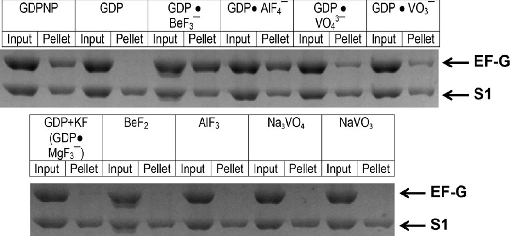
Figure 1.
Binding of EF-G to vacant ribosomes in the presence of various nucleotides and phosphate analogues (as indicated) measured by the pelleting assay. EF-G was incubated with vacant ribosomes. Half of each sample was pelleted through a sucrose cushion (pellet); the other half was used as a loading control (input). Protein content of ribosome pellets was analyzed using SDS-PAGE. The band corresponding to EF-G and the largest ribosomal protein S1 are indicated by arrows.

