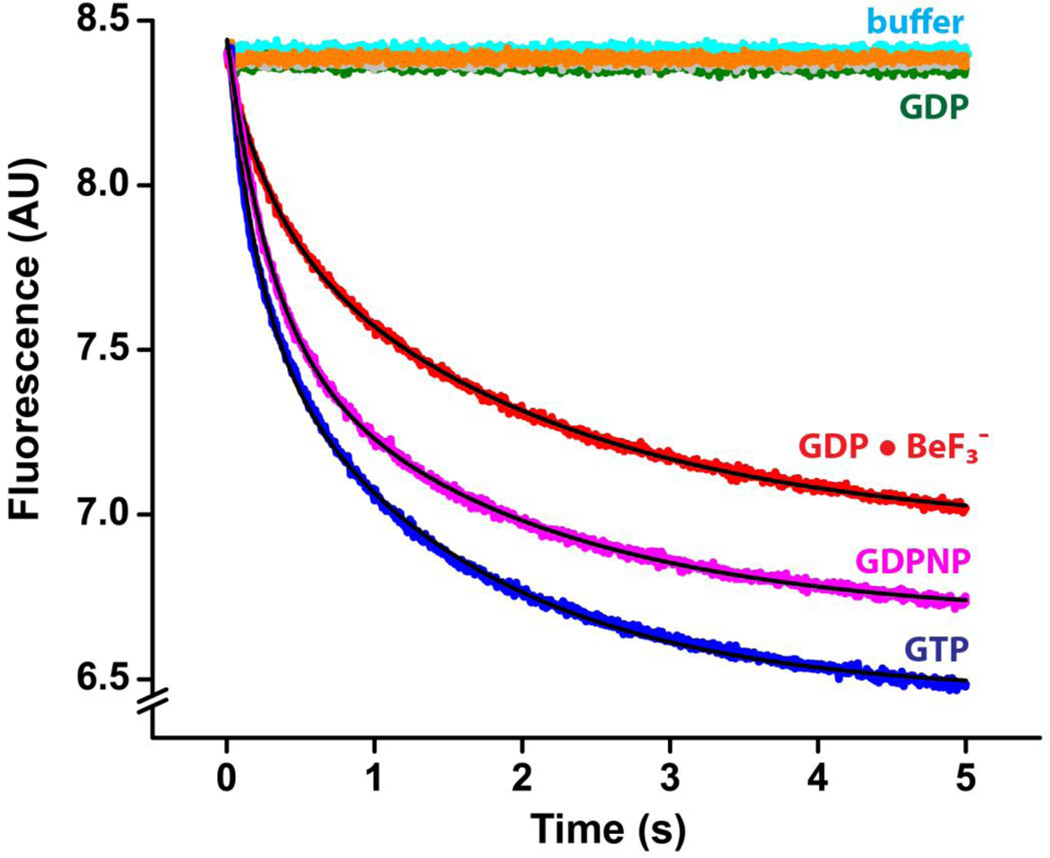
Figure 3.
Pre-steady-state kinetics of translocation in the presence of phosphate analogues at pH 7.5. mRNA translocation was induced by mixing pretranslocation ribosomes (35 nM after mixing) with EF-G (1 µM after mixing) preincubated with GTP (blue), GDPNP (magenta), GDP (dark green), GDP and KF (grey), GDP and VO43− (orange) or GDP and BeF3− (red). Experiments were performed in polyamine buffer at pH 7.5. mRNA translocation was detected by the quenching of fluorescein attached to the 3’ end of mRNA using a stopped-flow apparatus. Pretranslocation ribosomes were also mixed with buffer only (cyan) to account for the rates of the photobleaching of fluorescein and spontaneous translocation. Double-exponential fits for fluorescence quenching (black lines) are reported in Table 1.

