FIGURE 7.
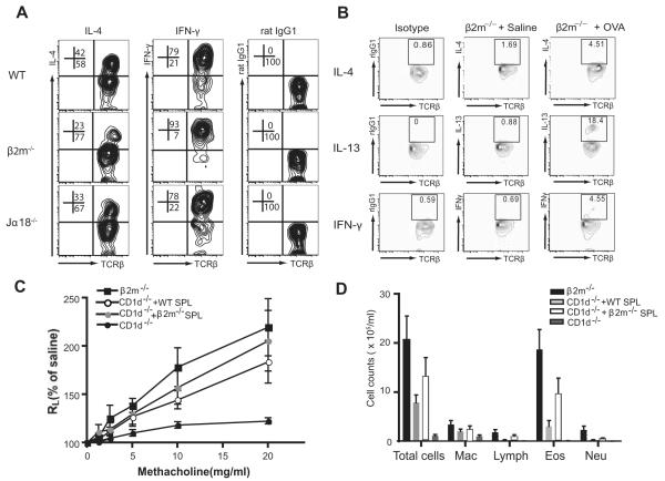
Cytokine production in NKT cells from β2m−/− mice. A, NKT cells in β2m−/− mice produce IL-4 cytokine. Spleen cells were isolated as in Fig. 6A, stimulated with PMA and ionomycin, and stained with anti-NK1.1 mAb, anti-TCRβ mAb, and mAbs against IL-4 or IFN-γ, which are representative of three experiments. The numbers are shown as a percentage of NK1.1+ T cells. B, Spontaneous cytokines production from lung NKT cells of OVA-immunized β2m−/− mice. Lung cells were obtained 24 h after the last OVA or saline challenge in β2m−/− mice. Lymphocytes were enriched, cultured for 3 h without further stimulation, and stained with anti-NK1.1 mAb, anti-TCRβ mAb, and mAbs against IL-4, IL-13, or IFN-γ. The numbers shown represent the percentage of NK1.1+ T cells after gating on NK1.1+ cells. Lung NKT cells from OVA-immunized β2m−/− mice produced more IL-13, but not much IL-4 and IFN-γ, compared with saline-treated β2m−/− mice. C, Adoptive transfer of a NKT cell-enriched population from β2m−/− mice rescues AHR in NKT cell-deficient CD1d−/− mice. In brief, 2 × 10⁷ spleen cells enriched for NKT cells from WT or β2m−/− mice were adoptively transferred into OVA-sensitized CD1d−/− mice. The recipients were then challenged with OVA intranasally and analyzed for AHR. D, BAL fluid was taken from the mice described in C and evaluated for cell type. See Fig. 1 legend for definitions of abbreviations.

