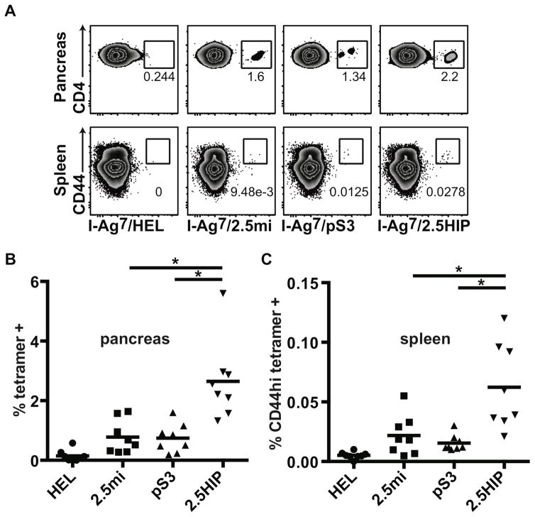
Fig. 3. Tetramer analysis of pancreas and spleen of diabetic NOD mice.
Single cell suspensions were prepared from pancreas and spleen of diabetic NOD female mice (n = 8) and stained with tetramers, antibodies and a live cell marker (7AAD). Gates were set on live leukocytes (7AAD-, CD45), CD4, dump- (CD8, CD11b, CD11c, CD19, F4/80) cells. (A) Tetramer staining in the pancreas and the spleen of a representative mouse. (B) Summary of CD4 tetramer-positive cells present in the pancreas. (C) Summary of CD4 CD44hi tetramer-positive cells present in the spleen. Each symbol represents a different mouse. Averages are indicated as a black horizontal bar. Data are from 4 independent experiments and were analyzed by two-tailed unpaired t-test. Statistical significance (*) was defined at a p value <0.05.

