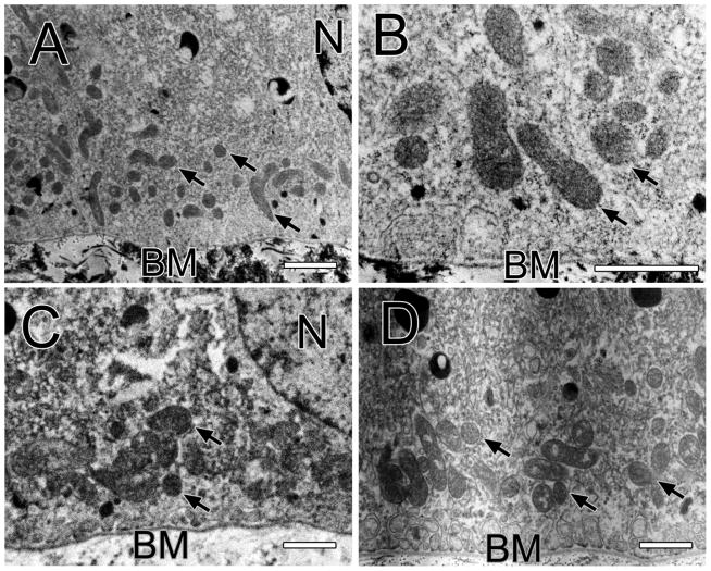
Figure 1.
Figure 1A shows a macula RPE cell of the one-year old monkey with the basal membrane (BM) below and the apical side above as in all electron micrographs. Arrows point to mitochondria and a nucleus (N) is labeled. Bar, 1 μm in all panels. Figure 1B shows a magnified view of a macular RPE cell of the one-year old monkey. Several mitochondria are labeled with arrows. Bruch’s membrane (BM) is labeled. Figure 1C shows a macular RPE cell of the two-year old monkey. Many mitochondria (arrows) are located between the nucleus (N) and the basal membrane (BM) of the cell. Figure 1D shows the basal and middle third of a RPE cell of the 6.5-year old monkey. A group of mitochondria (arrows) are located just above the basal membrane.

