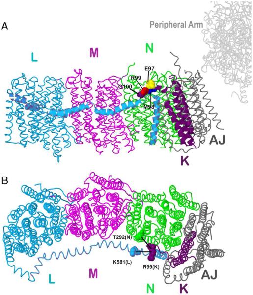
Fig. 1.
Schematic view of the membrane arm of Complex I showing the sites of cross-linking and stop codon substitutions. Subunit L is colored blue, subunit M magenta, subunit N green, subunit K purple and subunits A and J are colored gray. A. Residues G100 (purple), R99 (red), M98 (blue) and E97 (yellow) are showed in space-filling. B. Two cross-links between subunit K and N or L are also indicated. The image was developed from Protein Data Bank file 3rko (16). The view is from the cytoplasm. The peripheral arm would be to the right. The crystal form of this membrane arm from E. coli is lacking subunit H, but overall its structure is very similar to that of the entire Complex I structure determined from Thermusthermophilus (PDB file 4hea) (Baradaran et al. 2013).

