Fig. 2.
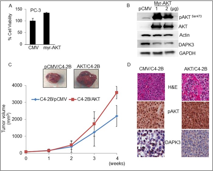
AKT negatively regulates DAPK3 in CaP cells. (A) PC-3 cells were transiently transfected with myr-AKT or empty vector (CMV) and subjected to MTT assay. (B) PC-3 cells were transiently transfected with myr-AKT and empty vector (CMV), and cell lysates were prepared for western blot analysis for pAKT, AKT, and DAPK3 proteins. β-actin or GAPDH was used as a loading control. (C) For xenograft studies, C4-2B/pCMV or C4-2B/AKT cells in a final volume (1.5 × 10⁶/50 µl ) were injected subcutaneously in the flanks of mice. The mice were monitored twice weekly, and tumor volumes were measured once a week for 4 weeks. A line graph represents the tumor growth and volume (mm3) of C4-2B/pCMV and C4-2B/AKT tumors. (D) Xenograft tumors were analyzed for H&E as well as IHC for pAKT and DAPK3.

