
Figure 1.
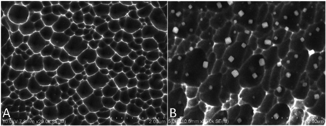
SEM images (20,000×) of the surface of bare metal stent (A) and anti-CD34 antibodies modified stent (B). The SEM results revealed that the pore size was 384.1 ± 160.6 nm wide and 127.7 ± 49.4 nm deep. SEM, scanning electron microscope.
Official websites use .gov
A
.gov website belongs to an official
government organization in the United States.
Secure .gov websites use HTTPS
A lock (
) or https:// means you've safely
connected to the .gov website. Share sensitive
information only on official, secure websites.
SEM images (20,000×) of the surface of bare metal stent (A) and anti-CD34 antibodies modified stent (B). The SEM results revealed that the pore size was 384.1 ± 160.6 nm wide and 127.7 ± 49.4 nm deep. SEM, scanning electron microscope.