
Figure 4.
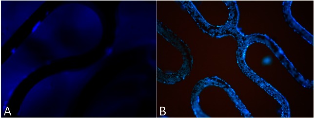
Fluorescence microscopic images (40×) of CD34+ KG-1a cells stained with DAPI on bare metal stent (A) or anti-CD34 antibodies modified stent (B). Vast numbers of cells (63,500 ± 6,500 cells/mm2) of CD34+ positive KG-1a cells were captured on the stent surface with CAMs, whereas only a small number of KG-1a cells were observed on the BMS surface. BMS, bare metal stent; CAMs, anti-CD34 antibody-modified stent; DAPI, 4’-6-diamidino-2-phenylindole.