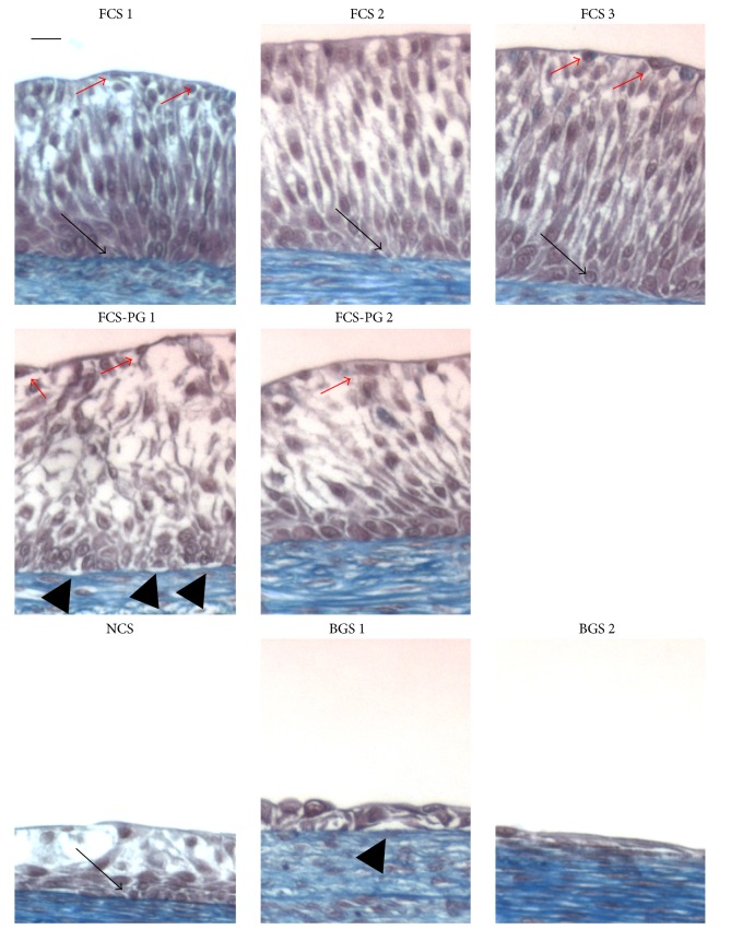
Figure 3.
Uroepithelium differentiation was better when FCS was used for cell culture compared to the use of postnatal sera. Masson's trichrome staining of tissues produced using human Fb and UC. Scale bar = 10 μm. Black arrows point to basal lamina (dark blue line after Masson's trichrome staining). Red arrows point clear superficial cells (umbrella cells). Black arrowheads point to detachment of the epithelium from the basal lamina.

