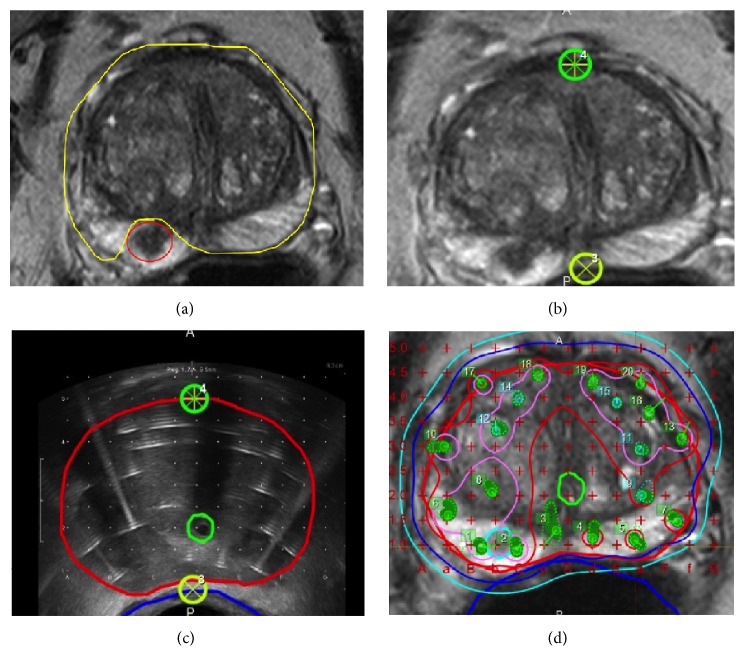
Figure 3.
(a) Preprocedural MRI demonstrating GTV (yellow) and nondiseased prostate (red). Preprocedural MRI using two anchor points (green and yellow circles) (b) are able to be registered to intraprocedural TRUS with 2.5 mm grid spacing (c). (d) The resulting merge of the TRUS grid and preprocedural MRI with GTV (thick light blue), urethra (thick solid light green), rectum (thick blue line), and isodose lines (thin lines) for typical whole gland plan using 18 HDR catheters.

