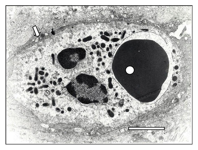
Figure 18.

Transmission electron microscopy of a RBC phagocytosed (◯) by a polymorphonuclear cell-neutrophil (⇒) in the liver of a sheep infected with T. vivax at 15 days. Notice the RBC inside a phagocytic vacuole surrounded by lysosomes. Bar = 1.5 μm.
