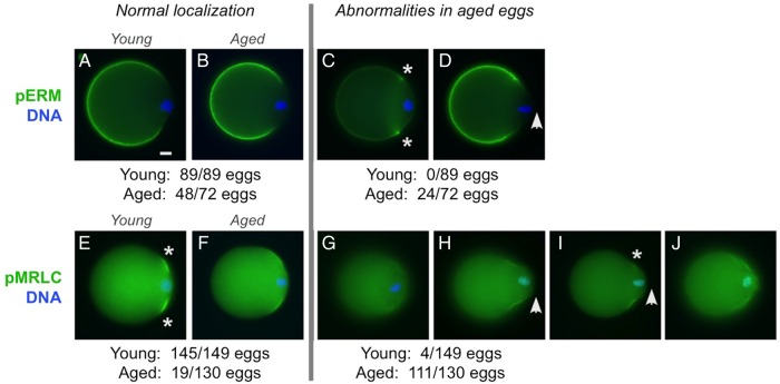
Figure 2.
pERM and pMRLC localizations in post-ovulatory aged metaphase II eggs. Immunofluorescence analysis of pERM and pMRLC in young (A and E) and aged (B–D and F–J) eggs. (Note: These studies focused on metaphase II young and aged eggs; parthenogenetically activated aged eggs are not included here.) Microvillar pERM was observed in young eggs (89/89; A) and the majority of aged eggs (48/72; B). Abnormalities in pERM localization and cell morphology were observed in 24 out of 72 aged eggs, such as enriched pERM localization at the junction between the amicrovillar and microvillar domains (asterisks in C), or the presence a protruding amicrovillar domain (arrowhead in D). Normal localization of pMRLC in the amicrovillar domain was observed in most young eggs (145/149; E), but fewer aged eggs [19/130 (P < 0.0001); F]. Reduced pMRLC signal in the amicrovillar domain was observed in 4 out of 149 young eggs and 30 out of 130 aged eggs (G). Aged eggs showed other various pMRLC abnormalities, including a protruding amicrovillar domain (H, I; arrowhead), uneven pMRLC distribution around the perimeter of the amicrovillar domain (I; asterisk marking the side of the amicrovillar domain with fainter pMRLC), and patchy pMRLC staining with a wrinkled appearance in the amicrovillar domain (J). Scale bar (in A), 10 µm.

