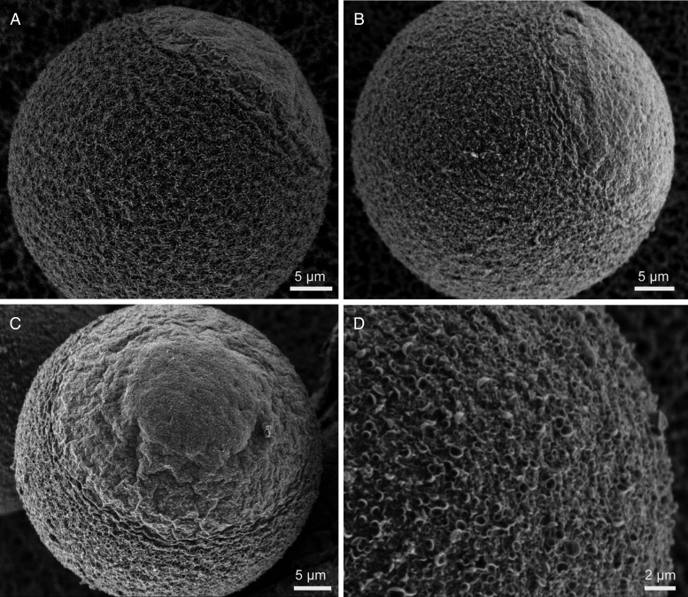
Figure 5.
Membrane topography in control and ML-7-treated young, unfertilized eggs. LVSEM was used to examine control eggs (A) and ML-7-treated fertilized eggs (B–D). As shown previously (Eager et al., 1976; Nicosia et al., 1978; Longo and Chen, 1985), control eggs have two distinct surface domains, the microvillar and amicrovillar domains. In ML-7-treated eggs, the amicrovillar domain either seemed mostly normal (B; 4/7 eggs for which we had a sufficient view of the amicrovillar domain), or was somewhat distended (Panel C; 3/7 eggs). The microvillar domain of 17 out of 19 ML-7-treated eggs analyzed showed varying extents of crater-like structures on the egg surface, shown in higher magnification in D (see also Fig. 6 for these features in fertilized ML-7-treated eggs). Scale bars equal 5 µm in A–C, and 2 µm in D.

