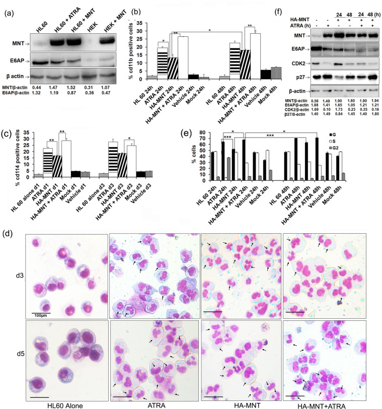
Figure 4. MNT overexpression promotes differentiation of HL60 cells.
a. HL60 cells were transiently transfected with 1.0μg HA-MNT and WCEs was prepared post-48h transfection. Lysates were resolved on 8% SDS-PAGE followed by immunoblotting with anti-MNT, E6AP and β-actin antibodies. b. Graphical representation of FACS analysis of cd11b in HL60 cells transiently transfected with HA-MNT (1.0μg) and co-treated with 1μM ATRA for 24 and 48h. c. Graphical representation of FACS analysis of cd114 in HL60 cells transiently transfected with HA-MNT (1.0μg) and co-treated with 1μM ATRA post 24h transfection. d. HA-MNT overexpression leads to granulocytic differentiation in HL60 cells. Cells were stained with May-Grunwald and Wright's Giemsa staining. Arrows indicate granulocytes/neutrophils. Scale represent 100μm. e. Graphical representation of percentage of G0/G1 cells post HA-MNT (1.0μg) transfection and co-treatment with ATRA post-36h transfection. f. HA-MNT over expressing HL60 cells were immunoblotted with antibodies against indicated proteins. Data are representative of minimum three independent experiments. Results are given as standard error of mean (+S.E.M.); *P<0.05, **P<0.001, ***P<0.0001. One-way ANOVA with Bonferroni's Multiple Comparison Test was performed using GraphPad Prism Version 5.00.

