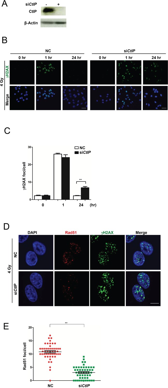
Figure 2. Loss of CtIP causes HRR deficiency.

A. Western blot analysis of CtIP in whole cell extracts from MCF7 cells transfected with CtIP or control siRNA (25 nM) for 48 hrs. B. The images of γH2AX foci after 4 Gy IR in control (NC) and CtIP-depleted MCF7 cells at different time points as indicated. Scale bar, 40 μm. C. Quantification of γH2AX foci in Figure 2B. Numbers of γH2AX foci were quantified from triplicated experiments (>50 cells at each condition) and are shown as mean values ± SEM. Statistical significance was calculated by one-way analysis of variance (ANOVA). ( * for P<0.05; ** for P<0.01; where not indicated, the P value was equal or higher than 0.05). D. Wild-type and CtIP-depleted MCF7 cells were irradiated (4 Gy) and fixed 3 hrs later. Rad51 and γH2AX foci were immunodetected with anti-Rad51 and anti-γH2AX antibodies, respectively. Cell nuclei were counterstained with DAPI. Scale bar, 10 μm. E. Quantification of Rad51 foci in Figure 2D. 50 cells at each condition were calculated. Mean ± SEM. Statisitcal significance, ** for P<0.01.
