
Figure 8. Inhibition of PI3K/Akt pathway promotes lysosome-mediated integrin degradation.
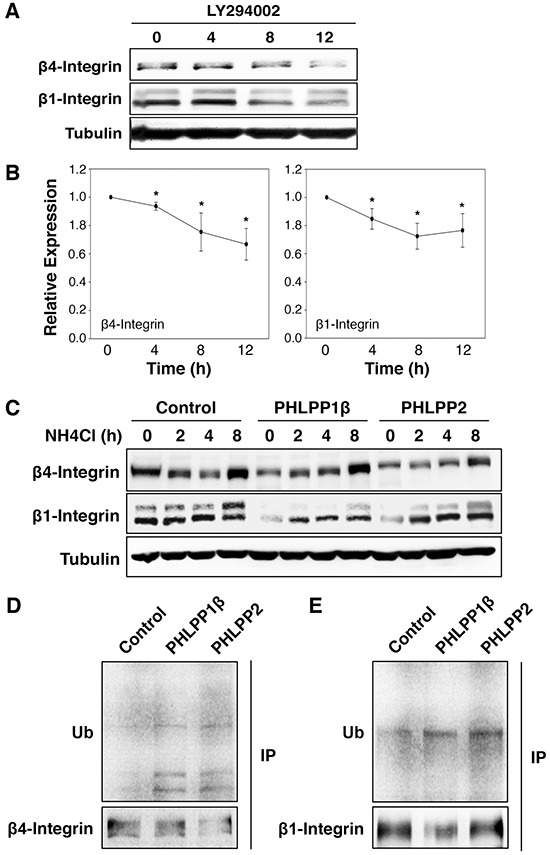
A. ASPC-1 cells were incubated with LY294002 for 0–12 hours. At indicated time points, cell lysates were prepared and analyzed for the expression of β1 and β4 integrin and tubulin using immunoblotting. B. Relative expression levels of β1 and β4 integrin at different time points following LY294002 treatment in ASPC-1 cells were calculated and normalized to tubulin. The level in untreated cells was set to 1. Data represent the mean ± SEM (n = 3, * p<0.05 by two-sample t-tests). C. Stable control and PHLPP overexpressing Panc-1 cells were treated with NH4Cl (5 mM) for 2, 4, and 8 hours. Cell lysates were prepared and analyzed by immunoblotting for integrin expression. D. Cell lysates prepared from stable control and PHLPP overexpressing Panc-1 cells were immunoprecipitated with antibodies against β1 or β4 integrin. The ubiquitination of endogenous β1 and β4 integrin was detected using the anti-ubiquitin antibody.