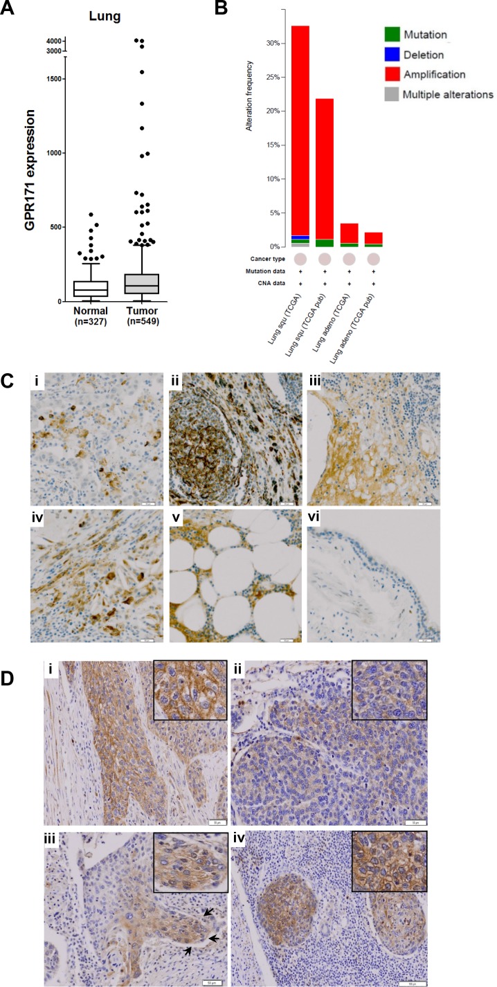
Figure 1. GPR171 expression is induced in lung cancer tissues.
A. GPR171 mRNA expression in normal lung and lung cancer tissues. Expression was assessed using the GENT database [38]. Boxes indicate the 75th percentile, median, and 25th percentile. Dots indicate outliers. B. Alteration frequency of GPR171 in lung squamous cell carcinoma (Lung squ) and adenocarcinoma (Lung adeno). C. Immunohistochemical analysis of GPR171 in lung cancer tissues.(i) Squamous cell carcinoma of the lung; (ii) adenocarcinoma of the lung; (iii) small-cell lung carcinoma; (iv) large-cell lung carcinoma; (v) lymph nodal metastatic carcinoma from adenocarcinoma of the lung; (vi) normal bronchial epithelium. Scale bars = 50 μm. D. Immunohistochemical analysis of GPR171 in (i) well-differentiated squamous cell carcinoma. Scale bars = 50 μm; (ii) poorly-differentiated squamous cell carcinoma. Scale bars = 50 μm; (iii) the invading front of squamous cell carcinoma. Arrows indicate invading tumor fronts in squamous cell carcinoma. Scale bars = 100 μm; (iv) lymph nodal metastatic tumor cells. Scale bars = 100 μm. Insets show magnified images to present GPR171 is expressed in the cytoplasm and cytoplasmic membrane.

