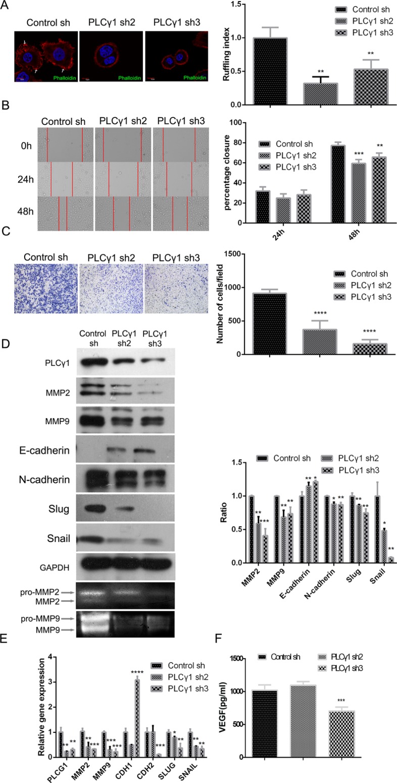
Figure 2. Lentivirus-mediated PLCγ1 shRNA could suppress migration in human gastric adenocarcinoma BGC-823 cells.

Cells were transduced with lentivirus-mediated PLCγ1 shRNA2/3 vectors. (A) The formation of membrane ruffles was detected using Ruffling assay as described in Materials and Methods. The cell nuclei were stained DAPI (blue) and the membrane ruffles were stained rhodamine-conjugated phalloidin (red). Scale bar = 10 μm. (B and C) The migration ability was measured using Transwell assay (B, magnification × 100) and Scratch assay (C, magnification × 400) as described in Materials and Methods. (D) The protein levels of MMP2, MMP9, E-cadherin, N-cadherin, snail, slug, and GAPDH were detected with Western blotting analysis, and the pro and active forms of MMP2/9 were observed using gelatin zymography assay as described in Materials and Methods. (E) The mRNA levels of PLCG1, MMP2, MMP9, CDHI, CDH2, SNAIL, SLUG, and GAPDH were detected using Real-time PCR analysis as described in Materials and Methods. (F) The level of VEGF in extracellular matrix was detected using ELISA as described in Materials and Methods. Data are reported as means ± S.D. of three independent experiments (*P < 0.05, **P < 0.01, ***P < 0.001, ****P < 0.0001, vs respective control).
