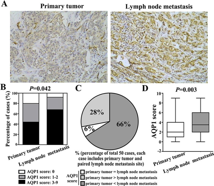
Figure 3. Cytoplasmic expression of AQP1 in lymph node metastases was higher than their paired primary tumors in total 50 paired cases.

(A) Representative immunohistochemical images of AQP1 expression in primary breast cancer and paired lymph node metastasis (magnification 200 ×). (B) 68% (34/50) lymph node metastasis specimens exhibited high AQP1 cytoplasmic expression, while 44% (22/50) primary breast cancer specimens showed high AQP1 cytoplasmic expression (χ2 = 6.343, P = 0.042). (C) Among total 50 paired cases (each case including primary tumor and paired lymph node metastasis specimens), 66% (33/50) cases showed that AQP1 expression in lymph node metastases was higher than paired primary tumors, 6% (3/50) cases showed that AQP1 expression in lymph node metastases was similar to their paired primary tumors and 28% (14/50) cases showed that AQP1 expression in lymph node metastasis was lower than their paired primary tumor. (D) Cytoplasmic AQP1 expression in primary breast cancer specimens (median score: 2.0) was lower than that in their paired lymph node metastases (median score: 3.5) (Mann-Whitney U test, P = 0.003).
