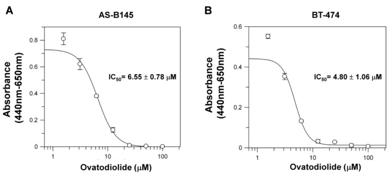
Figure 1.
The cytotoxic effect of ovatodiolide in human breast cancer cells. AS-B145 (A) or BT-474 (B) cells were seeded in a 96-well plate and treated with a different concentration of ovatodiolide (0, 1.625, 3.125, 6.25, 12.5, 25, 50, 100 μM) for 72 h (n = 4 for each concentration). Cell proliferation was determined by WST-1 reagent and the IC50 value was calculated by GraFit software. The experiments were repeated two times and results from a representative experiment were presented.

