Figure 5.
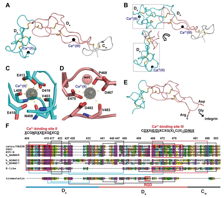
Arm structure. (A) The Ds, Da, and Cw subdomains of catrocollastatin/VAP2B (2DW0) are shown in cyan, pink, and gray, respectively. (B) The D domain of K-like proteinase (3K7N) with two different views of the Ds subdomain (in dotted line boxes). Close up views of the Ca2+-binding sites, II (C) and III (D) in catrocollastatin/VAP2B. (E) Structure of an RGD-containing disintegrin, trimestatin (1J2L). Suggested integrin-binding residues are indicated. (F) Amino acid sequence alignment of catrocollastatin/VAP2B (PDB: 2DW0_A), VAP1 (PDB: 2ERO_A), RVV-X (PDB: 2E3X_A), human ADAM28 (Genbank ID (GI): 98985828), human ADAM10 (GI: 29337031), human ADAM17 (GI: 14423632), K-like (PDB: 3K7Y_A) and trimestatin (1J2L_A) generated using Clustal X2 (http://www.clustal.org/clustal2/). Disulfide bonds and the boundaries of the subdomains are schematically indicated. Ca2+-binding sites II and III are boxed in red.

