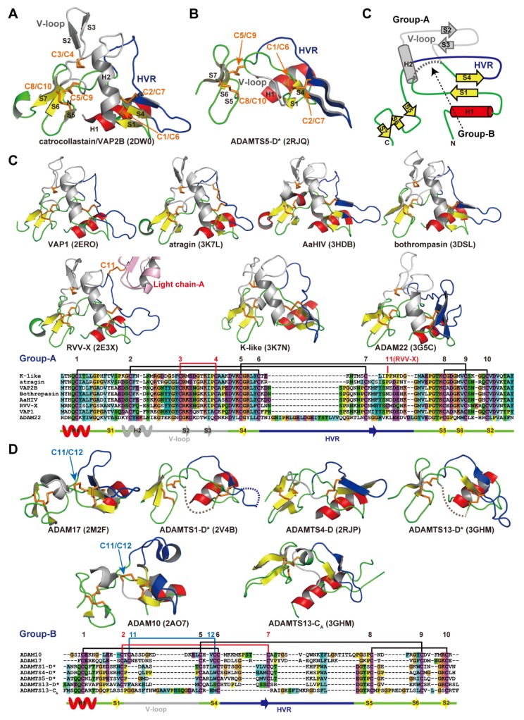
Figure 6.
ADAM_CR domain. Ribbon representation of the Ch subdomains of catrocollastatin/VAP2B (A) and the D* domain of ADAMTS5 (B). (C) Topology diagram of the ADAM_CR domain. Gallery of the group-A (C) and group-B (D) ADAM_CR domain structures. Conserved α-helix and β-strands are shown in red and yellow, respectively. Disulfide bonds, residues in HVR, and the residues in the V-loop are shown in orange, blue, and gray, respectively. The PDB ID for each protein structure is indicated in parentheses.

