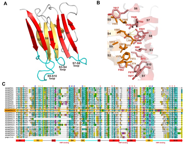
Figure 10.
S domain structure. (A) Ribbon representation of the crystal structure of the S domain of ADAMTS13. The strands in the two β-sheets are shown in red and orange. (B) Close-up view of the hydrophobic core between the β-sheets. Side chains forming the hydrophobic core and the conserved Glu641, whose side-chain oxygen atoms make hydrogen bonds with the backbone nitrogen atom of Leu595 in the opposing strand, are indicated. (C) Sequence alignment of the S domain of human ADAMTSs, ADAMTS-L and papilin. The residues in the hydrophobic core and the conserved aromatic surface cluster [53] are marked with * and #, respectively.

