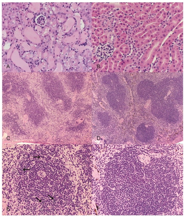
Figure 3.
Representative images from mice treated with orellanine (OR) or saline controls (CON). Hematoxylin and eosin. (A) (top left) Kidney from an OR-treated mouse revealing variable tubular degeneration and ectasia with luminal accumulations of proteinic fluid and occasional sloughed cells; (B) (top right) kidney from a CON mouse with normal tubular morphology; (C) (middle left) spleen from an OR-treated mouse revealing an overall reduction in red and white pulp cellularity; (D) (middle right) spleen from a CON mouse with normal cellularity and well-defined red and white pulp margins; (E) (bottom left) higher magnification of white pulp from an OR-treated mouse revealing numerous pyknotic and karyorrhectic lymphocytes (arrows); (F) (bottom right) higher magnification of white pulp from a CON mouse with normal morphology. Scales: A,B,E,F = 600× magnification; C,D = 100× magnification.

