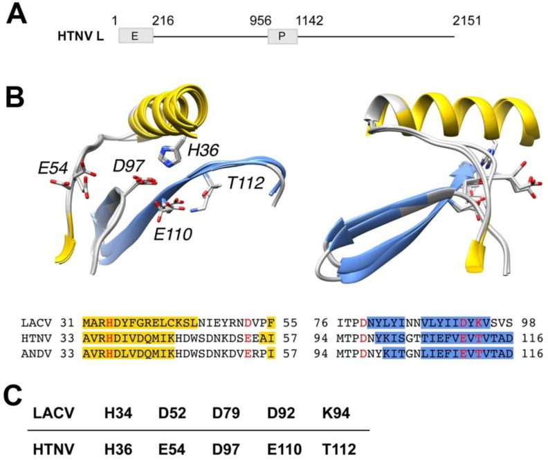
Figure 1.
Model of the putative active sites of prototypic Hantaan virus (HTNV) and Andes (ANDV) superposed on the active site of orthobunyavirus La Crosse (LACV). (A) Schematic representation of HTNV L protein. The L segment of the prototypic HTNV strain 76/118 has 6533 nucleotides and encodes the L protein of 2151 amino acids (aa). HTNV L contains a polymerase (P) domain (aa 956–1142) and a putative endonuclease (E) domain (aa 1–216). (B) Models of the putative active sites of HTNV and ANDV superposed on the active site of LACV. Ribbon diagrams of LACV (PDB entry 2XI5), HTNV and ANDV after structural superposition of key active site residues. Secondary structure classifications was calculated using definition of secondary structure proteins (DSSP) [33] for LACV or predicted using the method/program PSI-PRED [34]. Helix residues have been colored yellow and β-strand residues have been colored blue in the ribbon diagrams as well as in the alignment, according to DSSP or PSI-PRED results, respectively. Annotations in ribbon diagrams follow the HTNV residue numbering in the alignment. Key residues are colored red in the alignment and shown with side chains in the ribbon diagrams. The ribbon diagrams to the right have been rotated 90 degrees around the vertical coordinate axis. (C) Amino acid residues of the active site of HTNV L endonuclease derived from the model in (A).

