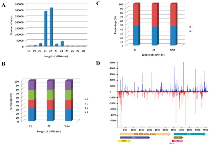
Figure 4.
Characterization of the MaYMV vsiRNAs. (A) Size distribution of the vsiRNAs along MaYMV genome; (B) the relative frequency of the four different 5′ terminal nucleotides in vsiRNAs; (C) polarity distribution of vsiRNAs that perfectly matched either plus or minus MaYMV genomic sequence; and (D) distribution profile of the vsiRNAs that revealed multiple hotspots along the MaYMV genome.

