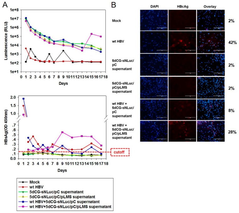
Figure 6.
Infection of primary tupaia hepatocytes (PTH) by 5dCG-sNLuc. (A) Analysis of NanoLuc (top) and HBsAg (bottom) in infected PTH supernatants. Freshly prepared PTH were infected with wild type HBV and/or transfected Huh-7 cell supernatants as indicated. Culture media were changed and collected at indicated time points, and assayed for NanoLuc activity and HBsAg; (B) Immunofluorescent analysis of HBcAg expression in infected PTH. Infected cells were fixed at day 17 post infection and intracellular HBcAg was detected using anti-HBcAg (Dako). Percentages of HBcAg-positive cells were calculated from ten randomly selected views and listed on the right. Cell nuclei were stained using DAPI. Scale bars, 200 µm.

