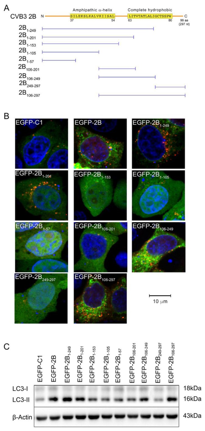
Figure 3.
The autophagy-inducing motif of protein 2B. (A) The diagram shows the constructs expressing truncated protein 2B fused with EGFP (not included in the plot); (B) HeLa cells were co-transfected with pEGFP-2BXX (representing the truncated 2B) and pmCherry-LC3 for 42 h and treated with E64-D/PEPA (10 ng/mL) for 6 h. The control cells were transfected with pEGFP-C1 and pmCherry-LC3. Cells were observed by confocal microscope (×600); (C) HeLa cells were treated as described in (B). EGFP and LC3 were detected by Western blotting. n = 4. Experiment was repeated three times. Representative images were presented.

