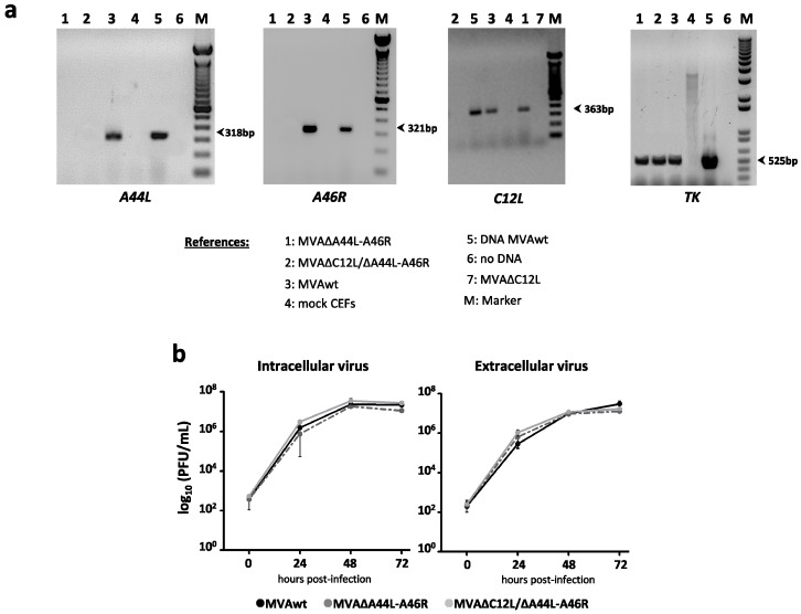
Figure 1.
In vitro characterization of MVA∆A44L-A46R and MVA∆C12L/∆A44L-A46R. (a) RNA was extracted from mock or infected CEFs with the different MVAs at 24 hpi (moi = 1), specific mRNAs were assessed by RT-PCR; and (b) virus growth kinetics of MVA vectors. BHK-21 were infected with the indicated MVA at moi = 0.01. At different hpi virus production was titrated by immunostaining.

