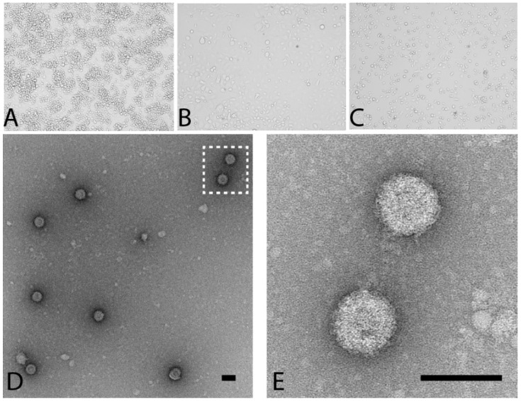
Figure 1.
Parry’s Lagoon virus (PLV) Morphology and Growth Characteristics. PLV induces distinct CPE on C6/36 monolayers at 5 days post infection. (A) mock-infected C6/36 cells; (B) PLV-infected C6/36 cells and (C) Corriparta virus (CORV)-infected C6/36 cells; (D,E) Culture supernatant of C6/36 cells infected with PLV was concentrated and virions negatively stained using a 1% uranyl acetate solution. Scale bar represents 80 nm.

