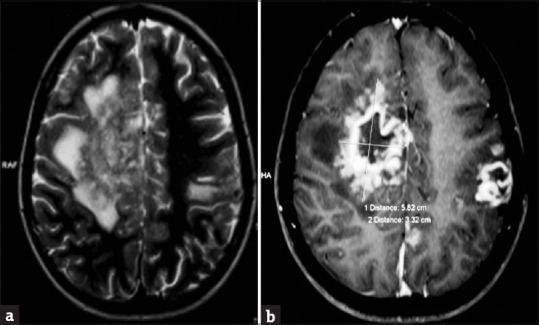
Figure 6.

(a) Axial T2 and flair image demonstrating an iso- to hypointense mass lesion in the right frontal lobe with a peripheral sun burst pattern and perilesional edema. Left frontparietal hyperintensity is also noted. (b) Axial T1-weighted image revealing rim enhancing lesions in bilateral frontal lobes with sunburst/gyral pattern in the periphery surrounded by a rim of hypointensity producing a “double ring sign”
