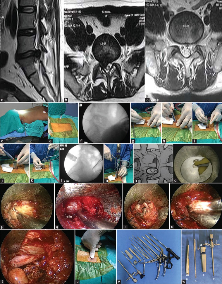
Figure 1.
(a) Sagittal magnetic resonance imaging T2W lumbosacral spine showing prolapsed intervertebral disc (PIVD) at L5S1 level. (b) Axial magnetic resonance imaging T2W showing PIVD L5-S1. (c) Postoperative axial magnetic resonance imaging. (d) Patient positioning. (e) Localization with 18 G spinal needle. (f) Confirmation of correct placement by IITV image. (g) Skin and fascial incision. (h) Dilatation with Arthrospine dilator. (i) Sliding of arthrospine tube over dilator. (j) Dilator is withdrawn and Arthrospine tube is held in place. (k) Arthrospine working insert with scope and sheath is snug fit over Arthrospine tube. (l) IITV Confirmation of correct placement of Arthrospine tube. (m) Nibbling of superior lamina to gain entry into canal. (n) Laminotomy diagrammatic. (o) Endoscopic view of interlaminar window on mannequin. (p) Endoscopic view of interlaminar window. (q) View of endoscopic laminotomy. (r) Endoscopic view of extruded disc. (s) Disc removal by disc forceps. (t) Endoscopic view of decompressed nerve root. (u) Incision after subcuticular closure. (v) Arthrospine assisted discectomy instrumentation. (w) Arthrospine system. (a) Arthrospine dilator, (b) Dural and nerve root retractor, (c) Arthrospine tube, (d) Arthrospine working insert, (e) Arthrpscope sheath

