Fig. 3.
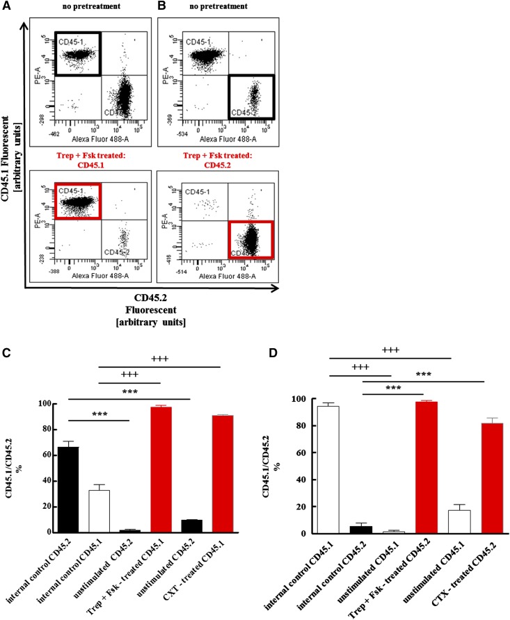
Competitive advantage of HSPCs after pretreatment with the combination of treprostinil and forskolin or cholera toxin over unstimulated controls. Murine HSPCs were isolated from CD45.1+ and from CD45.2+ donor mice. Either CD45.1+ (A, C) or CD45.2+ (B, D) cells were incubated for 1 hour in medium in the absence of any additional stimulus, in the presence of the combination of 10 μM treprostinil and 30 μM forskolin (Trep + Fsk, pretreated) or of 10 μg ml−1 cholera toxin (as a positive control). First, cell suspensions were prepared that contained either equivalent amounts of both unstimulated CD45.1+ and CD45.2+ cells [top panel in (A); bars labeled “internal control” in (C)] or equivalent amounts of stimulated and untreated donor cells [bottom panel in (A)]. These suspensions containing a total of 106 cells were administered by tail vein injection to lethally irradiated recipients (A, C). Conversely, CD45.2+ cells, which were either not stimulated [top panel in (B); bars labeled “internal control” in (D)], or stimulated [bottom panel in (B)], and untreated CD45.1+ were mixed at a ratio of 1:10; a total of 5 × 10⁵ cells were injected into recipient mice (B, D). The proportion of bone marrow cells arising from the two sources was determined by flow cytometry 16 weeks after transplantation. Representative original dot plots are shown in (A) and (B); bar diagrams in (C) and (D) represent the means ± S.E.M. (n = 5 animals per group) from two independent experiments. The indicated statistical comparisons of CD45.2- (+) and CD45.1-positive cells (*) were done by analysis of variance (ANOVA) followed by Tukey’s multiple comparison (P = 0.0001).

