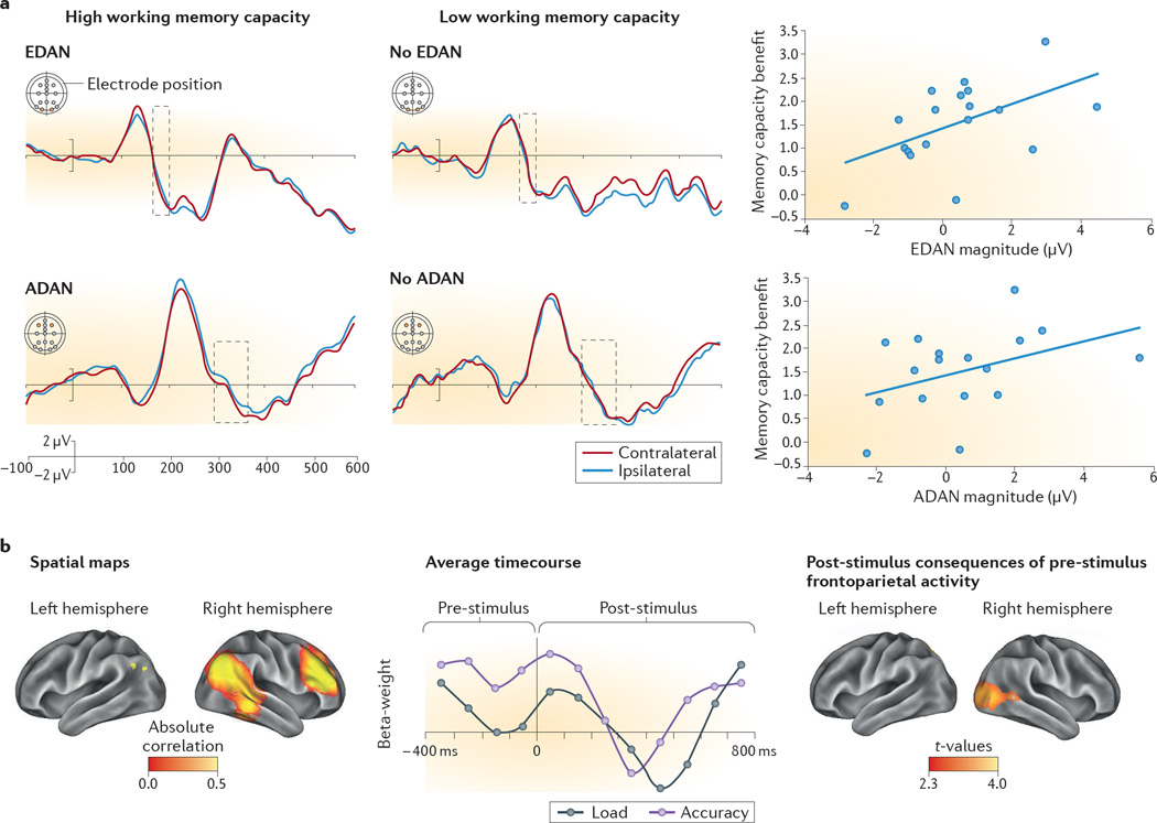
Figure 2. Visual attention correlates with working memory capacity.
Electrophysiological and magnetoencephalographic studies provide evidence that variation in the neural markers of attentional deployment correlate with individual differences in memory capacity. a | Electrophysiological markers of visuo-spatial attentional orienting in preparation for encoding information into memory distinguish 10-year-old children with higher or lower working memory capacity. The early directing attention negativity (EDAN) is an event-related potential locked to the onset of spatial cues that direct attention, and it is characterized by greater negativity at posterior scalp electrodes that are contralateral) than at posterior scalp electrodes that are ipsilateral to the direction of the attention-orienting cue. EDAN is thought to indicate cue-processing. Another event-related potential, anterior directing attention negativity (ADAN), is also characterized by greater negativity at scalp electrodes contralateral to cue direction, but the electrodes used for this analysis are more anterior than those used for EDAN. ADAN is associated with deployment of attentional control. The waveforms (left-hand and central panels) represent the average time course of these differences for children with high working memory capacity (who do show EDAN and ADAN); and children with low working memory capacity (who do not show EDAN and ADAN). The area marked with a dashed box highlights when the waveforms differ for contralateral and ipsilateral sites between children with low and high working memory capacity. The scatterplots (right-hand panels) show significant correlations between the magnitudes of EDAN and ADAN and the benefits of cues for memory on this task. b | Magnetoencephalographic data suggest that the preparatory oscillations of a right frontoparietal network before encoding items into memory predict the accuracy of later memory recall and the activity of visual cortices when the memoranda are first encoded. Adults and 10-year-old children were asked to encode either two (low load) or four (high load) simultaneously presented items into memory and then to recall whether a probe item was among the memoranda. The children’s spatial maps of right-frontoparietal network oscillations are shown on the left and the time course of the effect of these oscillations on memory accuracy are shown in the centre. Represented on the x-axis is time, with 0 indicating the time point at which the to-be-encoded items were presented. The y-axis represents beta weights. Pre-stimulus activity in this frontoparietal network in preparation of encoding successfully discriminates trials in which participants remember items accurately (purple line). By contrast, this frontoparietal network is not differentially engaged by memory load (an index of task difficulty; grey line). The panel on the right represents the area in the children’s visual cortex whose activity after the onset of the to-be-encoded stimuli was significantly predicted by right frontoparietal network activity, illustrating the coupling that occurs between this network and the visual cortex. Part a is reprinted with permission from REF. 91, Massachusetts Institute of Technology. Part b is reprinted from REF 90.

