
Fig. 2.
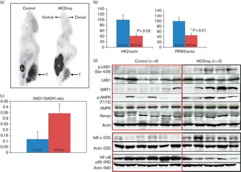
MCDmp-induced alterations in energy-dependent signaling pathways. (a) 18F-FDG PET scan shows decreased 18F-FDG uptake within tumors in the MCDmp group. T, tumor, b, urinary bladder. (b) The expression of the key enzymes for glycolysis (HK2 and PKM2) decreased significantly in the MCDmp group. (c) NAD+/NADH ratio was significantly increased (>3-fold) in melanoma tissues removed from the MCDmp group compared with the control group (*P=0.044, n=15 in each group). (d) Western-blot analysis showed activation of the LKB1/SIRT1/AMPK loop in the MCDmp group. There was no significant alteration in Nampt expression. Therefore, the NAD+/NADH ratio could be increased in the MCDmp group, independent of Nampt expression. In addition, the MCDmp intervention suppressed NF-κB p65 expression in the nuclear fraction of cells along with increased cytosolic IkB.