
Fig. 3.
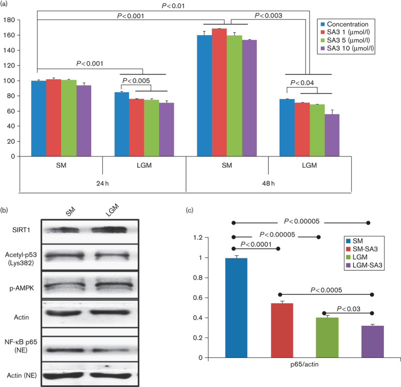
Tumor-suppressive role of SIRT1 under an energy restriction condition. (a) MTT assay of B16F10 melanoma cells cultured in a standard medium (SM, 11 mmol/l of glucose in RPMI-1640 medium) showed no significant cell proliferation at 24 or 48 h after treatment of SA3. However, cell proliferation was significantly suppressed after SA3 treatment when melanoma cells were culture in a low-glucose medium (LGM, 5.5 mmol/l of glucose). (b) SIRT1 and p-AMPK were increased in melanoma cells cultured in an LGM. However, the acetyl-p53 level was not significantly decreased despite an increased SIRT1 level. NF-κB p65 in the nuclear fraction was decreased under a low-glucose condition. (c) NF-κB level was decreased after SA3 treatment under both standard and low-glucose conditions.