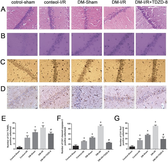
FIGURE 1.
Cerebral injury induced by hindlimb ischemia–reperfusion is aggravated by diabetes. (A) Representative images of brain tissue from the hippocampal CA1 area taken from rats in the control-sham, control-I/R, T1DM-sham, T1DM-I/R and DM-I/R+TDZD-8 groups, showing (A) H&E staining, (B) TUNEL staining, (C) immunohistochemical staining of cleaved caspase-3 and (D) immunohistochemical staining of Iba1 (magnification, 200×). (E–G) Quantitative analysis of the data shown in (B–D), respectively. *P < 0.05 versus the control-sham group; #P < 0.05 versus the DM-I/R group.

